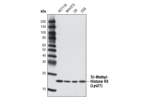
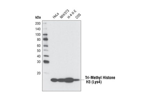
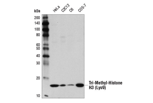
For optimal ChIP and ChIP-seq results, use 10 μl of antibody and 10 μg of chromatin (approximately 4 x 10⁶ cells) per IP. This antibody has been validated using SimpleChIP® Enzymatic Chromatin IP Kits.
The CUT&RUN dilution was determined using CUT&RUN Assay Kit #86652.
Application
Dilution
Western Blotting
1:1000
Immunohistochemistry (Paraffin)
1:50 - 1:200
Immunofluorescence (Immunocytochemistry)
1:25600 - 1:102400
Flow Cytometry (Fixed/Permeabilized)
1:50 - 1:200
Chromatin IP
1:50
Chromatin IP-seq
1:50
CUT&RUN
1:50
Storage
Supplied in 10 mM sodium HEPES (pH 7.5), 150 mM NaCl, 100 µg/ml BSA, 50% glycerol and less than 0.02% sodium azide. Store at –20°C. Do not aliquot the antibody.
For a carrier free (BSA and azide free) version of this product see product #53775.
For western blots, incubate membrane with diluted primary antibody in 5% w/v BSA, 1X TBS, 0.1% Tween® 20 at 4°C with gentle shaking, overnight.
NOTE: Please refer to primary antibody product webpage for recommended antibody dilution.
A. Solutions and Reagents
From sample preparation to detection, the reagents you need for your Western Blot are now in one convenient kit: #12957 Western Blotting Application Solutions Kit
NOTE: Prepare solutions with reverse osmosis deionized (RODI) or equivalent grade water.
20X Phosphate Buffered Saline (PBS): (#9808) To prepare 1 L 1X PBS: add 50 ml 20X PBS to 950 ml dH2O, mix.
10X Tris Buffered Saline (TBS): (#12498) To prepare 1 L 1X TBS: add 100 ml 10X to 900 ml dH2O, mix.
1X SDS Sample Buffer: Blue Loading Pack (#7722) or Red Loading Pack (#7723) Prepare fresh 3X reducing loading buffer by adding 1/10 volume 30X DTT to 1 volume of 3X SDS loading buffer. Dilute to 1X with dH2O.
10X Tris-Glycine SDS Running Buffer: (#4050) To prepare 1 L 1X running buffer: add 100 ml 10X running buffer to 900 ml dH2O, mix.
10X Tris-Glycine Transfer Buffer: (#12539) To prepare 1 L 1X Transfer Buffer: add 100 ml 10X Transfer Buffer to 200 ml methanol + 700 ml dH2O, mix.
10X Tris Buffered Saline with Tween® 20 (TBST): (#9997) To prepare 1 L 1X TBST: add 100 ml 10X TBST to 900 ml dH2O, mix.
Treat cells by adding fresh media containing regulator for desired time.
Aspirate media from cultures; wash cells with 1X PBS; aspirate.
Lyse cells by adding 1X SDS sample buffer (100 µl per well of 6-well plate or 500 µl for a 10 cm diameter plate). Immediately scrape the cells off the plate and transfer the extract to a microcentrifuge tube. Keep on ice.
Sonicate for 10–15 sec to complete cell lysis and shear DNA (to reduce sample viscosity).
Heat a 20 µl sample to 95–100°C for 5 min; cool on ice.
Microcentrifuge for 5 min.
Load 20 µl onto SDS-PAGE gel (10 cm x 10 cm).
NOTE: Loading of prestained molecular weight markers (#59329, 10 µl/lane) to verify electrotransfer and biotinylated protein ladder (#7727, 10 µl/lane) to determine molecular weights are recommended.
Electrotransfer to nitrocellulose membrane (#12369).
C. Membrane Blocking and Antibody Incubations
NOTE: Volumes are for 10 cm x 10 cm (100 cm2) of membrane; for different sized membranes, adjust volumes accordingly.
I. Membrane Blocking
(Optional) After transfer, wash nitrocellulose membrane with 25 ml TBS for 5 min at room temperature.
Incubate membrane in 25 ml of blocking buffer for 1 hr at room temperature.
Wash three times for 5 min each with 15 ml of TBST.
II. Primary Antibody Incubation
Incubate membrane and primary antibody (at the appropriate dilution and diluent as recommended in the product webpage) in 10 ml primary antibody dilution buffer with gentle agitation overnight at 4°C.
Wash three times for 5 min each with 15 ml of TBST.
Incubate membrane with Anti-rabbit IgG, HRP-linked Antibody (#7074 at 1:2000) and anti-biotin, HRP-linked Antibody (#7075 at 1:1000–1:3000) to detect biotinylated protein markers in 10 ml of blocking buffer with gentle agitation for 1 hr at room temperature.
Wash three times for 5 min each with 15 ml of TBST.
Proceed with detection (Section D).
D. Detection of Proteins
Directions for Use:
Wash membrane-bound HRP (antibody conjugate) three times for 5 minutes in TBST.
Prepare 1X SignalFire™ ECL Reagent (#6883) by diluting one part 2X Reagent A and one part 2X Reagent B (e.g. for 10 ml, add 5 ml Reagent A and 5 ml Reagent B). Mix well.
Incubate substrate with membrane for 1 minute, remove excess solution (membrane remains wet), wrap in plastic and expose to X-ray film.